Abstract
Objectives: The purpose of this study was to investigate the change in the posture of dental hygiene students and clinical dental hygienists when implementing dental scaling before and after posture correction training using the rapid upper limb assessment (RULA) method and 3D motion analysis. Methods: Thirty-two healthy volunteers performed dental scaling to remove artificial calculus on dental manikin. The movement and angle of the joints were verified by RULA and 3D motion analysis during the procedure. The subjects were also photographed for 1 minute during the procedure for 10 minutes while the calculus was removed. After the removal of the calculus, the subject and the instructor checked the video together. Posture correction training was conducted by the instructor so that the subject could perform the calculus removal operation in the correct posture. Artificial calculus of the adjacent teeth was then removed for the same period of time, and the change in posture was reviewed. Results: The total score of the posture change using RULA was 5.72 ± 0.58 before training and 4.31 ± 0.10 after training, showing a significant decrease after training (p<0.001), and upper arm, lower arm, wrist position, neck and waist position showed significant decrease after training. The three-dimensional motion analysis showed significant differences according to the criteria measured at all measurement sites except the left shoulder (p<0.05) Conclusions: It was confirmed through RULA and 3D motion analysis that postural correction training using calculus removal images was effective, and that correct postural education is essential to preventing musculoskeletal diseases caused by removal of calculus.
Figures & Tables
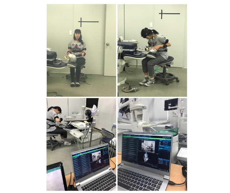
Fig. 1. Posture of dental calculus removal and 3D motion analysis program